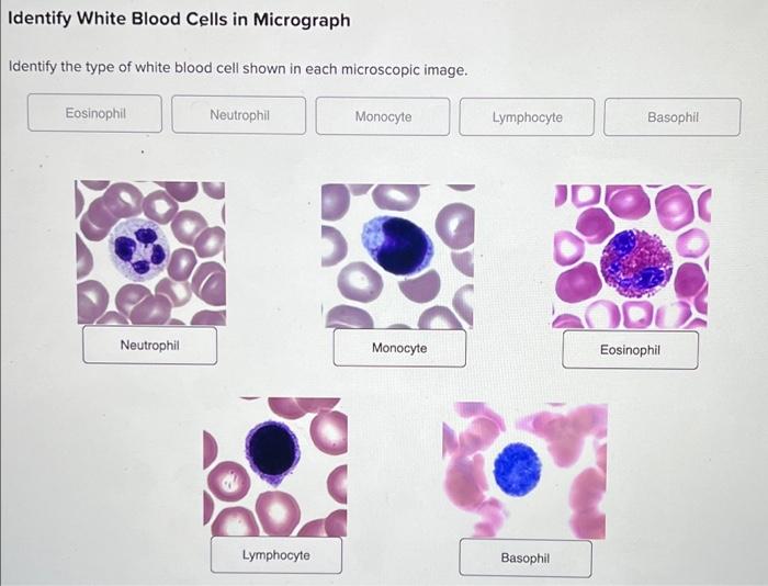
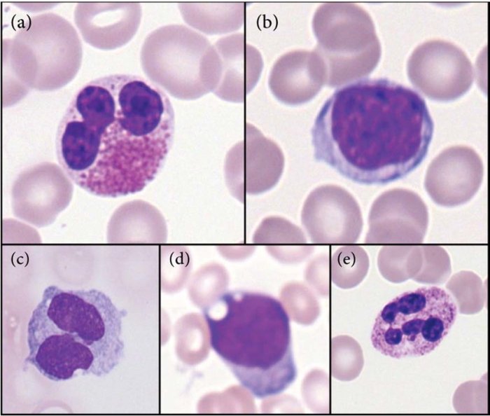
Identify the leukocytes shown in the photomicrographs below – As we delve into the identification of leukocytes presented in photomicrographs, this introductory paragraph invites readers into a realm of scientific exploration, where knowledge is meticulously dissected and presented with clarity and precision. Prepare to embark on an intellectual journey that promises to illuminate the intricacies of leukocyte identification.
The subsequent paragraphs will meticulously describe the distinct types of leukocytes, their morphological characteristics, and their crucial roles within the immune system. We will also explore the clinical significance of leukocyte identification, examining how it aids in diagnosing and monitoring various diseases.
A case study will be presented to illustrate the practical application of leukocyte identification in clinical settings.
Leukocyte Identification: Identify The Leukocytes Shown In The Photomicrographs Below

Photomicrographs provide a valuable tool for identifying leukocytes, which are essential components of the immune system. Leukocytes are classified into different types based on their morphology, staining characteristics, and functional roles. Understanding the key characteristics of each leukocyte type is crucial for accurate identification and interpretation of photomicrographs.
Morphology and Function, Identify the leukocytes shown in the photomicrographs below
The morphological features of leukocytes can provide important clues to their identity. Neutrophils are characterized by their multi-lobed nucleus and abundant cytoplasmic granules. Lymphocytes have a large, round nucleus and scant cytoplasm. Monocytes are larger than lymphocytes and have a horseshoe-shaped nucleus.
Eosinophils have a bilobed nucleus and eosinophilic granules in their cytoplasm. Basophils have a multi-lobed nucleus and basophilic granules in their cytoplasm.
Each leukocyte type plays a specific role in the immune system. Neutrophils are phagocytic cells that engulf and destroy bacteria. Lymphocytes are responsible for adaptive immunity, recognizing and eliminating specific pathogens. Monocytes differentiate into macrophages, which are phagocytic cells that remove cellular debris and pathogens.
Eosinophils are involved in defending against parasitic infections. Basophils release histamine and other inflammatory mediators.
Clinical Significance
Identifying different leukocytes in photomicrographs is clinically significant as it can aid in diagnosing and monitoring various diseases. For example, an elevated neutrophil count may indicate an infection, while an increased lymphocyte count may suggest a viral infection. Monocytosis can be a sign of chronic inflammation or certain types of leukemia.
Eosinophilia is associated with allergic reactions and parasitic infections. Basophilia can be a sign of allergic reactions or certain types of leukemia.
Case Study Analysis
In a clinical case, a patient presented with fever, cough, and shortness of breath. A photomicrograph of a blood smear revealed an increased number of neutrophils and lymphocytes. This finding, along with the patient’s symptoms, suggested a bacterial pneumonia. Further tests confirmed the presence of Streptococcus pneumoniae in the patient’s sputum, leading to a diagnosis of bacterial pneumonia.
Technical Considerations
The techniques used to prepare and stain photomicrographs for leukocyte identification involve blood smears and staining methods. Blood smears are prepared by spreading a thin layer of blood onto a glass slide. The slides are then stained using various techniques, such as Wright-Giemsa stain, which differentially colors the different leukocyte types.
Limitations and potential errors associated with leukocyte identification in photomicrographs include misidentification due to overlapping morphological features, artifacts from the staining process, and the presence of immature or atypical cells.
Educational Resources
- Textbook:Wintrobe’s Clinical Hematology
- Article:“Leukocyte Identification in Photomicrographs” by the American Society for Clinical Pathology
- Online Resource:“Leukocyte Identification Tutorial” by the University of Utah
Continuing education in this field is essential to stay updated on the latest advancements in leukocyte identification and interpretation, as well as to ensure accurate diagnosis and patient management.
Commonly Asked Questions
What are the different types of leukocytes?
Leukocytes are broadly classified into two main groups: myeloid and lymphoid cells. Myeloid cells include neutrophils, eosinophils, basophils, and monocytes, while lymphoid cells encompass lymphocytes (T cells, B cells, and natural killer cells).
How can leukocytes be identified in photomicrographs?
Leukocytes can be identified based on their size, shape, nuclear morphology, and cytoplasmic features. Specific staining techniques, such as Wright-Giemsa stain, can further enhance their visualization and differentiation.
What is the clinical significance of leukocyte identification?
Leukocyte identification plays a crucial role in diagnosing and monitoring various diseases, including infections, inflammatory conditions, hematological disorders, and immune deficiencies. Abnormal leukocyte counts or differentials can provide valuable clues to the underlying pathological processes.

